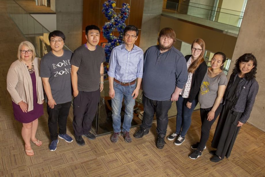
Yang
Laboratory
Skeletal Biology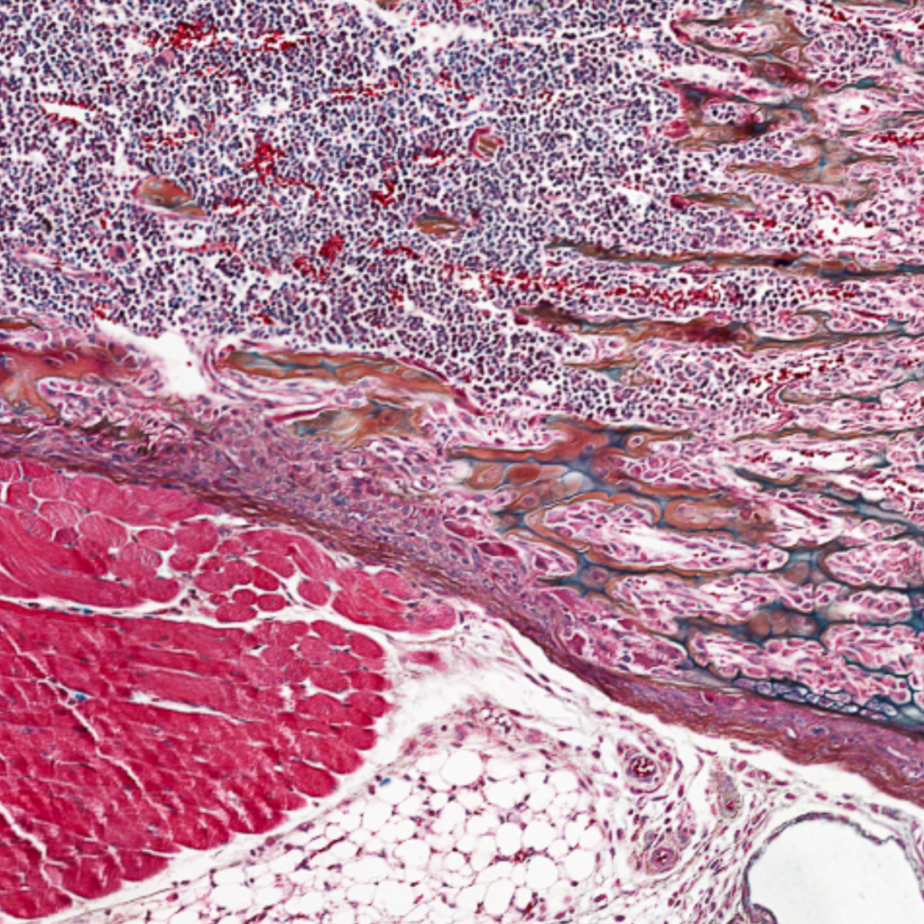
The Yang Lab identifies molecular mechanisms involved in skeletal development, stem cell fate determination, homeostasis and aging. We also design new gene and stem cell therapy approaches for skeletal tissue regeneration or disease treatments. To do this, we leverage skeletal disease models, comprehensive phenotyping, integrated multi-omics, and molecular and cellular biology methods.
Research Areas
SUMOylation is a branch of ubiquitination-like (Ubl) posttranslational modifications (PTMs) that conjugate monomeric SUMO (an ~100 aa protein tag) or polySUMO chains to target proteins, with a strong connection to stress responses and aging. A major function of mono sumoylation is to recruit sumo-interacting motifs (SIMs)-containing proteins to form protein complexes. In contrast, polysumoylation commonly destabilizes target proteins by promoting their ubiquitination, resulting in complex disassembly. Such versatility in protein complexes assembly/disassembly makes sumoylation important for cells to respond and adapt to changes and stresses. Additionally, sumoylation can mask other PTM sites or alter protein spatial conformation. SUMO modifications can be deconjugated by SENP family desumoylases, most of which remove only mono-sumoylation, whereas SENP6 and SENP7 can edit/remove poly-SUMO chains. The sumoylation/desumoylation equilibrium regulates many cellular processes, including DNA damage response, mitochondrial dynamics, epigenetic regulation, cell proliferation, senescence and apoptosis.
After SNPs in the SENP6 gene locus were identified as a top genetic risk factor for osteoarthritis, we began exploring the role of sumoylation in skeletal development and diseases. We found that losing SENP6 in mice leads to skeletal development defects and premature aging, partly due to elevated apoptosis and senescence in the progenitor compartments. Mechanistically, SENP6 desumoylates TRIM28, a multifaceted protein involved in chromatin silencing and p53 inhibition. Senp6 loss results in excessive SUMOylation of TRIM28; this weakens TRIM28’s function as a p53 suppressor, causing p53 hyperactivation and elevated senescence. Because chondrocyte senescence, joint aging and osteoarthritis (OA) susceptibility function in a feed-forward loop, SENP6 and its downstream pathways may be used as therapeutic targets. We are currently working in this direction. In addition, we are studying SENP6’s role in osteoporosis in various cellular contexts.
We also found that a sumoylation inhibitor, ginkgolide acid, overrides mesenchymal stem cells’ fate from osteoblast toward adipocyte, even under osteogenic differentiation conditions. Meanwhile, we have developed a tracking tool to dynamically visualize polysumoylated proteins in live cells, providing a new approach to the sumoylation research community.
As with other stem cells, lineage-specific transcription factors and the microenvironment cooperatively regulate skeletal stem cell (SSC) maintenance and differentiation, but their effectiveness depends on the permissiveness of the epigenetic landscape. The epigenetic landscape of a cell reflects its history of lineage progression and predetermines its future identity. In the mammalian skeletal system, appendicular bones and most axial bones develop from mesoderm-derived skeletal precursors/stem cells (SSCs). In contrast, most craniofacial bones arise from ectoderm-derived neural crest cells (NCCs). NCCs have a default neural/glial and epidermal fate but acquire an osteochondrogenic potential through transient reactivation of pluripotency. We were curious whether and how chromatin silencing marks contribute to establishing SSC identity and guide SSC lineage progression for skeletogenesis. TRIM28 is an adaptor protein that recruits specific enzymes to promote H3K9me3 and DNA methylation, which makes it ideal for addressing this question. Our study found that loss of TRIM28 in mesoderm-derived cells expands the SSC pool, but weakens SSC osteochondrogenic potential while endowing SSCs with properties of ectoderm-derived NCCs, leading to severe defects of skeletogenesis. TRIM28 preferentially silences chromatin regions that are more accessible in NCCs; loss of this silencing upregulates neural gene expression and enhances neurogenic potential. Moreover, TRIM28 loss causes hyperexpression of GREM1, an extracellular signaling factor promoting SSC self-renewal and neurogenic potential by activating AKT/mTORC1 signaling. This work suggests that TRIM28-mediated chromatin silencing establishes a barrier to securing the SSC lineage trajectory and prevents a transition to ectodermal fate by regulating intrinsic and microenvironment cues.
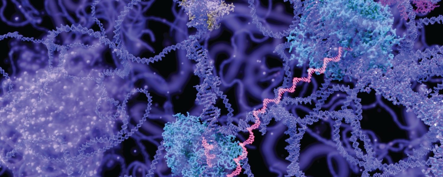
The other branch of our research focuses on how epigenetic mechanisms contribute to the development of age-related skeletal diseases. With age, cells gradually lose chromatin silencing marks, such as DNA methylation or histone methylation. Loss of these marks leads to the reactivation of transposable elements (TEs), which comprise about 50% of the human genome. Reciprocally, the host organism develops chromatin-silencing mechanisms to counteract/silence TE activity. Endogenous retroviruses (ERVs), a major type of TE, are genetic remnants of past retrovirus infections in the host genome throughout evolution. Currently, most ERV elements are not transposed. However, they still contribute to cis-regulatory function and genetic diversity in the hosts or produce double-stranded RNAs (dsRNAs) that stimulate inflammation response. Our lab found that the chromatin accessibility and transcription of ERVs, particularly from evolutionarily “intermediate age” ERV families, were enriched and elevated in OA cartilage. We also found that global H3K9me3 levels were reduced in osteoarthritic or aging cartilage. This study unveils the intricate relationship between epigenetic alterations, ERV activation and OA, paving the way for potential therapeutic interventions by targeting these pathogenic mechanisms. Our lab seeks a comprehensive understanding of the interaction between aging, ERV activation and OA pathogenesis, with the goal of using this knowledge to alleviate OA development.
In collaboration with the Krawczyk Lab at VAI, we are studying how an X-linked lysine demethylase governs sex-dimorphic bone mass regulation by modulating energy metabolism in osteoclast lineage. The outcome of this work may lead to new approaches to enhance women’s bone health.
We study signaling dysregulation and cellular stress-related skeletal disorders. We are also exploring innovative approaches to alleviate OA symptoms and regenerate articular cartilage in the OA joints through stem cell and gene therapies.
News & Featured Publications
Learn More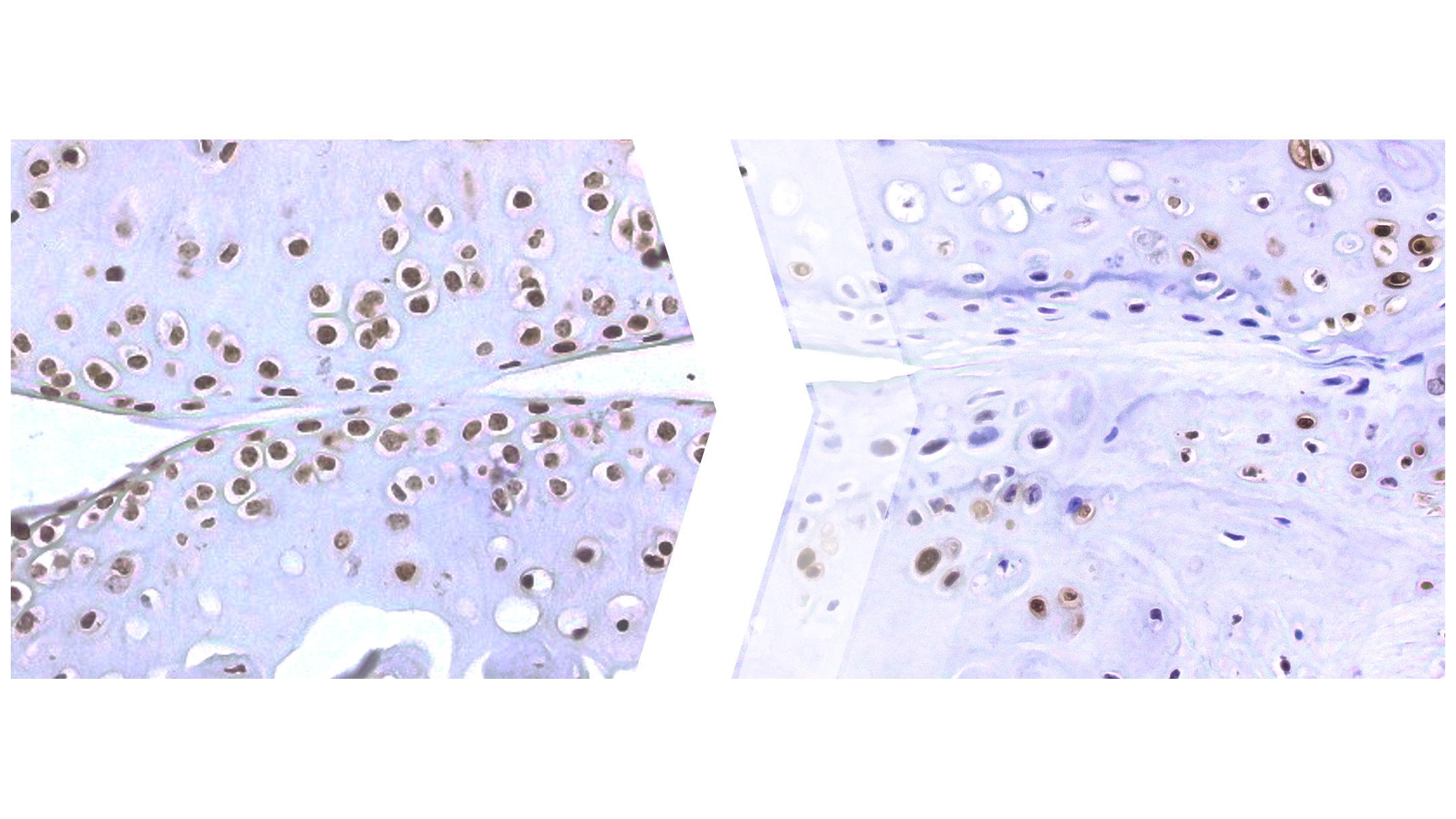
Could silencing old viruses help treat osteoarthritis?
‘A Grand Plan' for achieving breakthroughs and improving health

Generosity of VAI employees empowers osteoporosis discovery
Floramo JS, Molchanov V, Liu H, Liu Y, Craig SEL, Yang T. 2023. An integrated view of stressors as causative agents in OA pathogenesis. Biomolecules 13(5):721.
Liu H, Zhai L, Liu Y, Lu D, Vander Ark A, Yang T*, Krawczyk CM*. 2023. The histone demethylase KDM5C controls female bone mass by promoting energy metabolism in osteoclasts. Sci Adv 9(14).
*Co-corresponding authors
Liu H, Liu Y, Jin SG, Johnson J, Xuan H, Lu D, Li J, Zhai L, Li X, Zhao Y, Liu M, Craig SEL, Floramo JS, Molchanov V, Li J, Li JD, Krawczyk C, Shi X, Pfeifer GP, Yang T.2023. TRIM28 secures skeletal stem cell fate during skeletogenesis by silencing neural gene expression and repressing GREM1/AKT/mTOR signaling axis. Cell Rep 42(1):112012.
Our Impact
We’re raising thousands to save millions.
We’re turning hope into action for the millions of people around the world affected by diseases like cancer and Parkinson’s. Find out how you can help us make a difference.
- 141 peer-reviewed papers published in 2025, 74 of which were in high-impact journals
- 15 VAI-SU2C Epigenetics Dream Team clinical trials launched to date
- 10 clinical trials and related projects supported by VAI through the International Linked Clinical Trials Program
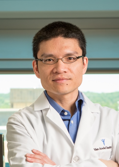
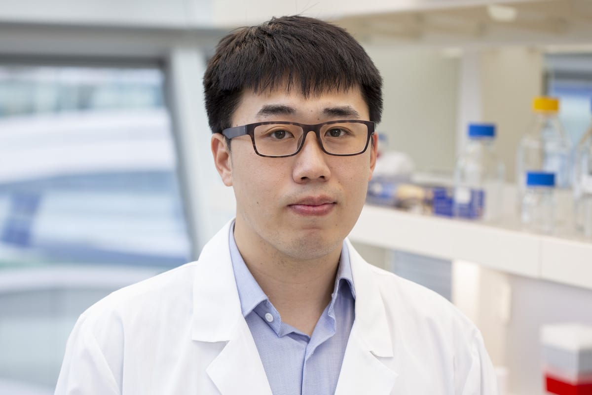
Tao Yang, Ph.D.
Professor, Department of Cell Biology
Areas of Expertise
Skeletal development and aging, osteoarthritis, osteoporosis, skeletal stem cells, sumoylation, signaling pathways
Biography
Dr. Yang received his B.S. in biochemistry from Wuhan University, and his Ph.D. in biochemistry from the Shanghai Institute of Biochemistry and Cell Biology, Chinese Academy of Sciences (SIBCB, CAS). He then joined Baylor College of Medicine as a postdoctoral fellow working with Drs. Paul Overbeek and Brendan Lee. In 2009, he began serving as an instructor in the Department of Molecular and Human Genetics at Baylor. Dr. Yang joined VAI as an assistant professor in February 2013.
SELECTED PUBLICATIONS
Liu Y, Molchanov V, Brass D, Yang T. 2025. Recent advances in omics and the integration of multi-omics in osteoarthritis research. Arthritis Res Ther 27(1):100.
Liu Y, Molchanov V, Zhao Y, Lu D, Liu H, Jang J, Yang T. 2025. H3K9me3 loss and ERVs activation as hallmarks for osteoarthritis progression and knee joint aging. Osteoarthritis Cartilage 33(1):128–133.
Floramo JS, Molchanov V, Liu H, Liu Y, Craig SEL, Yang T. 2023. An integrated view of stressors as causative agents in OA pathogenesis. Biomolecules 13(5):721.
Liu H, Zhai L, Liu Y, Lu D, Vander Ark A, Yang T*, Krawczyk CM*. 2023. The histone demethylase KDM5C controls female bone mass by promoting energy metabolism in osteoclasts. Sci Adv 9(14).
*Co-corresponding authors
Liu H, Liu Y, Jin SG, Johnson J, Xuan H, Lu D, Li J, Zhai L, Li X, Zhao Y, Liu M, Craig SEL, Floramo JS, Molchanov V, Li J, Li JD, Krawczyk C, Shi X, Pfeifer GP, Yang T. 2023. TRIM28 secures skeletal stem cell fate during skeletogenesis by silencing neural gene expression and repressing GREM1/AKT/mTOR signaling axis. Cell Rep 42(1):112012.
Liu H, Craig SEL, Molchanov V, Floramo JS, Zhao Y, Yang T. 2022. SUMOylation in skeletal development, homeostasis, and disease. Cells 11(17):2710.
Autosomal recessive LRP1-related syndrome featuring cardiopulmonary dysfunction, bone dysmorphology, and corneal clouding. Cold Spring Harb Mol Case Stud 8(6):a006169.
Yang CH*, Fagnocchi L*, Apostle S, Wegert V, Casani-Galdón S, Landgraf K, Panzeri I, Dror E, Heyne S, Wörpel T, Chandler DP, Lu D, Yang T, Gibbons E, Guerreiro R, Brás J, Thomasen M, Grunnert LG, Vaag AA, Gillberg L, Grundberg, E, Conesa A, Körner A, PERMUTE, Pospisilik JA. 2022. Independent phenotypic plasticity axes define distinct obesity subtypes. Nat Metab.
*Co-first authorship
**Highlighted in News & Views
Liu Y, Molchanov V, Yang T. 2021. Enzymatic machinery of ubiquitin and ubiquitin-like modification systems in chondrocyte homeostasis and osteoarthritis. Curr Rheumatol Rep 23:62.
Generation and characterization of mouse models for skeletal disease. Methods Mol Biol 2221:165–191.
Wang W, Wang M, Ruan Y, Tan J, Wang H, Yang T, Li J, Zhou Q. 2019. Ginkgolic acids impair mitochondrial function by decreasing mitochondrial biogenesis and promoting FUNDC1-dependent mitophagy. J Agric Food Chem 67(36):10097–10106.
He Y, Shi J, Nguyen QT, You E, Liu H, Ren X, Wu Z, Li J, Qiu W, Khoo SK, Yang T, Yi W, Sun F, Xi Z, Huang X, Melcher K, Min B, Xu HE. 2019. Development of highly potent glucocorticoids for steroid-resistant severe asthma. Proc Natl Acad Sci U S A 116(14):6932–6937.
Lu D, Li J, Liu H, Foxa GE, Weaver K, Li J, Williams BO, Yang T. 2018. LRP1 suppresses bone resorption in mice by inhibiting the RANKL-stimulated NFκB and p38 pathways during osteoclastogenesis. J Bone Miner Res.
Maupin K, Weaver K, Bergsma A, Christie C, Zhong Z, Yang T, Williams BO. 2018. Enhanced cortical bone expansion in Lgals3-deficient mice during aging. Bone Res 6(7).
Liu H, Li J Lu D, Li J, Liu M, Williams BO, Li J, Yang T. 2018. Ginkgolic acid, a sumoylation inhibitor, promotes adipocyte commitment but suppresses adipocyte terminal differentiation of mouse bone marrow stromal cells. Sci Rep 8:2545.
Li J, Lu D, Dou H, Liu H, Weaver K, Wang W, Li J, Yeh ETH, Williams BO, Zheng L, Yang T. 2018. Desumoylase SENP6 maintains osteochondroprogenitor homeostasis by suppressing the p53 pathway. Nat Commun. 9:143.
Yang T, Williams BO. 2017. Low-density lipoprotein receptor-related proteins in skeletal development and disease. Physiol Rev 97(3):1211-1228.
Li J, Lu D, Liu H, Williams BO, Overbeek P, Lee B, Zheng L, Yang T. 2017. Deficiency of SCLT1 causes cystic kidney by activating ERK and STAT3 signaling. Hum Mol Genet 26(15):2949–2960.
Grafe I, Alexander S, Yang T, Lietman C, Homan EP, Munivez E, Chen Y, Jiang MM, Bertin T, Dawson B, Asuncion F, Ke HZ, Ominsky MS, Lee B. 2016. Sclerostin antibody treatment improves the bone phenotype of Crtap-/- mice, a model of recessive Osteogenesis Imperfecta. J Bone Miner Res 31(5):1030–1040.
He Y, Shi J, Yi W, Ren X, Gao X, Li J, Wu N, Weaver K, Xie Q, Khoo SK, Yang T, Huang X, Melcher K, Xu HE. 2016. Discovery of a highly potent glucocorticoid for asthma treatment. Cell Disc doi:10.1038/celldisc.2015.35.
Lu L, Harutyunyan K, Jin W, Wu J, Yang T, Chen Y, Joeng KS, Bae Y, Tao J, Dawson BC, Jiang MM, Lee B, Wang LL. 2015. RECQL4 regulates p53 function in vivo during skeletogenesis. J Bone Miner Res 30(6):1077–1089
Chen S, Grover M, Sibai T, Black J, Rianon N, Rajagopal A, Munivez E, Bertin T, Dawson B, Chen Y, Jiang MM, Lee B, Yang T, Bae Y. 2015. Losartan increases bone mass and accelerates chondrocyte hypertrophy in developing skeleton. Mol Genet Metab 115(1):53–60.
Grafe I, Yang T, Alexander S, Homan EP, Lietman C, Jiang MM, Bertin T, Munivez E, Chen Y, Dawson B, Ishikawa Y, Weis MA, Sampath TK, Ambrose C, Eyre D, Bächinger HP, Lee B. 2014. Excessive transforming growth factor-β signaling is a common mechanism in osteogenesis imperfecta. Nat Med 20(6):670–675.
Yang T, Grafe I, Bae Y, Chen S, Chen Y, Bertin TK, Jiang M-M, Ambrose CG, Lee B. 2013. E-selectin ligand 1 regulates bone remodeling by limiting bioactive TGF-β in the bone microenvironment. Proc Natl Acad Sci USA 110(18):7336–7341.
Chen S, Tao J, Bae Y, Jiang MM, Bertin TK, Chen Y, Yang T, Lee B. 2012. Notch gain of function inhibits chondrocyte differentiation via Rbpj-dependent suppression of Sox9. J Bone Miner Res 28(3):649–659.
Bae Y, Yang T*, Zheng HC*, Campeau P, Chen Y, Bertin T, Dawson B, Munivez E, Tao J, Lee B. 2012. miRNA-34c regulates Notch signaling during bone developent. Hum Mol Genet 21(13):2991–3000.
*Contributed equally
Keller B, Yang T, Chen Y, Munivez E, Bertin TK, Zabel B, Lee B. 2011. Interaction of TGF- β and BMP signaling pathways during chondrogenesis. PLoS One 6(1):e16421.
Yang T, Mendoza RL, Lu H, Li K, Tao J, Shah R, Keller B, Jiang M-M, Chen Y, Bertin TK, Dabovic B, Engin F, Rifkin DB, Hicks J, Jamrich M, Beaudet AL, Lee B. 2010. E-Selectin Ligand 1 regulates growth plate homeostasis by inhibiting the intracellular processing and secretion of mature transforming growth factor β. J Clin Invest 120(7):2474–2485.
Tao J, Chen S, Yang T, Dawson B, Munivez E, Bertin T, Lee B. 2010. Osteosclerosis due to Notch gain of function is solely Rbpj-dependent. J Bone Miner Res 25(10):2175–2183.
Mao J, Yang T, Gu Z, Heird WC, Finegold MJ, Lee B, Wakil SJ. 2009. aP2-Cre-mediated inactivation of acetyl-CoA carboxylase 1 causes growth retardation and reduced lipid accumulation in adipose tissues under lipogenic conditions. Proc Natl Acad Sci USA 106(41):17576–17581.
Jiang M, Gao Y, Yang T, Zhu X, Chen J. 2009. Cyclin Y, a novel membrane-associated cyclin, interacts with PFTK1. FEBS Lett 583(13):2171–2178.
Engin F, Yao Z*, Yang T*, Zhou G, Bertin TK, Jiang MM, Chen Y, Wang L, Zheng H, Sutton RE, Boyce BF, Lee B. 2008. Dimorphic effects of Notch signaling in bone homeostasis. Nat Med 14(3):299–205.
*Contributed equally
Gao Y, Jiang M, Yang T, Ni J, Chen J. 2006. A Cdc2-related protein kinase hPFTAIRE1 from human brain interacting with 14-3-3 proteins. Cell Res 16(6):539–547.
Chen Q, Liang D, Yang T, Leone G, Overbeek PA. 2004. Distinct roles of individual E2Fs in induction of cell cycle entry in postmitotic lens fiber cells of transgenic mice. Dev Neurosci 26(506):435–445.
Yang T*, Liang D, Koch PJ, Hohl D, Kheradmand F, Overbeek PA*. 2004. Epidermal detachment, desmosomal dissociation and destabilization of corneodesmosin in Spink5-/- mice. Genes Dev 18(19):2354–2358.
*Corresponding authors
Yang T, Gao Y-K, Chen J-Y. 2002. KIAA0202, a human septin family member, interacting with hPFTAIRE1. Acta Biochem Biophys Sin 34(4):520–525.
Yang T, Chen J-Y. 2001. Identification and cellular localization of human PFTAIRE1. Gene 267(2):165–172.
Book chapters
Yang T, Gover M, Joeng K, Lee B. 2017. Fetal and neonatal bone development. Primer on the metabolic bone diseases and disorders of mineral metabolism. 9th ed. American Society for Bone and Mineral Research.
Yang T, Gover M, Joeng K, Lee B. 2013. Fetal and neonatal bone development. Primer on the Metabolic Bone Diseases and Disorders of Mineral Metabolism, 8th ed. Wiley-Blackwell.


Mu-Nung Hsu, Ph.D.
Research Scientist, Department of Cell Biology
Ye Liu, Ph.D.
Research Scientist, Department of Cell Biology
Jennifer Lorenz
Senior Administrative Assistant I, Department of Cell Biology

Di Lu, M.S.
Research Associate, Department of Cell Biology

